INTRODUCTION
Myotonic muscular dystrophy is an autosomal dominant disorder characterized by progressive weakness and myotonia, accompanied by multisystemic involvement [1]. It is the most common form of adult-onset muscular dystrophy. Two subtypes have been recognized, each harboring nucleotide expansion of noncoding regions.
Myotonic dystrophy type 1 (DM1) is caused by a large trinucleotide repeat expansion in the untranslated tail of the dystrophia myotonica protein kinase (DMPK) gene. This mutation leads to mis-splicing of mRNA species and results in abnormal cellular processes and diverse symptoms. Muscle weakness in DM1 patients begins from face, ankle dorsiflexor, and finger flexor, which progresses to the other skeletal muscles including respiratory muscle. Frequently accompanied are cardiac arrhythmia, cataract, diabetes mellitus, respiratory dysfunction, and cognitive impairment. Cardiac and respiratory dysfunction reduces median life span to the age of 55 years in DM1 patients. Most severe cases may show neonatal hypotonia with respiratory failure.
Myotonic dystrophy type 2 (DM2) is caused by unstable tetranucleotide repeat expansion (CCTG) in the first intron of the CNBP gene on chromosome 3q21. Like in DM1, the repeat expansion has been shown to cause mRNA splicing errors, which lead to widespread cellular abnormalities and multisystemic symptoms [2]. Unlike DM1, the extent of repeat expansion in DM2 is less related to the disease severity, and anticipation is not observed in DM2. Patients with DM2 show proximal muscle weakness and myalgia.
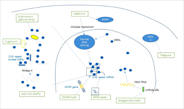
Current management of myotonic muscular dystrophy is focusing on symptomatic control of myotonia, cardiac care, and physical support of skeletal and respiratory muscle weakness. Multidisciplinary management by cardiologists, ophthalmologists, and endocrinologists for systemic complications is required. There is no cure or disease modifying therapy targeting the pathomechanism of myotonic muscular dystrophy. In this article, we briefly review molecular pathomechanism of DM1, and introduce emerging novel drugs in clinical development stages to treat DM1 (Fig. 1).
PATHOGENESIS OF MYOTONIC MUSCULAR DYSTROPHY
DM1 is caused by the expansion of the unstable CTG repeat in the 3´-untranslated region (UTR) of DMPK gene on chromosome 19q [1]. Normal individuals have 5 to 37 CTG repeats. Individuals with 38 to 49 CTG repeats are asymptomatic, but have risk of having children with large repeats by the phenomenon known as anticipation [3].
There are correlations between longer CTG repeat in DMPK and earlier age onset with more severe clinical manifestations. Patients with repeat expansions between 50 and 150 have lateonset, mild symptoms of DM1. CTG repeat ranges from 50 to 3,000 in patients with DM1. People with less than 70 repeat may not show neurologic symptom until their 6th decade, while those with 70-90 repeats may show mild symptoms after age 40. Congenital DM1 cases usually show more than 1,000 repeats [4]. The CTG repeat length varies widely by organ systems, which can make the correlation of repeat length and clinical severity less predictable. For example, CTG repeat is more stable in leukocytes than in skeletal or cardiac muscles, which can lead to underrepresentation of the result from blood tests compared with the burden of CTG repeat in other organ systems [5].
Expanded CTG repeat of DMPK gene transcribes into expanded CUG repeat on DMPK mRNA, which sequestraters RNA-binding proteins and disrupts splicing machinery. RNA-binding proteins such as muscleblind-like splicing regulator 1 (MBNL1) and CUG binding protein 1 (CUG-BP1) are affected [1]. MBNL1 co-locates in foci with the CUG repeat expansion [6]. Depleted MBNL1 results in abnormal splicing of mRNA transcription, which lead to abnormal expression of other genes [7]. For example, abnormal expression of chloride channel leads to myotonia, and loss of the insulin receptor proteins are associated with insulin resistance [8,9].
CUG repeat upregulates CUGBP1 proteins that also result in various abnormal mRNA splicing [10]. In addition, bi-directional antisense transcription, dysregulation of microRNAs and potentially non-ATG-mediated translation of homopolymeric toxic proteins have been introduced as novel mechanisms of DM1 [11].
DRUG DEVELOPMENT OF MYOTONIC MUSCULAR DYSTROPHY
At this point, there is no therapy that can cure myotonic dystrophy. Multidisciplinary management, involving neurology, cardiology, endocrinology, pulmonology and rehabilitation medicine, is needed to control widespread organ dysfunction. Recent advances in knowledge on the underlying molecular mechanism of myotonic dystrophy guides new approaches for disease specific treatments for DM1. Novel medications such as antisense oligonucleotides and gene therapies try to correct the disease process at the gene level. Small molecules to reduce toxic mRNA products or prevent interaction of CUG repeat and RNA-splicing proteins have been introduced. Some legacy drugs may be used for symptom controls or mitigate the metabolic derangement.
ANTISENSE OLIGONUCLEOTIDES (ASO)
ASOs are short, synthetic, single stranded oligonucleotides or its analogs that bind to a specific sequence of single strand RNA [12]. ASO therapies target the fundamental pathogenetic process, so it has theoretically higher chances of success than therapies targeting downstream pathways. Two distinctive strategies that use ASOs to treat myotonic dystrophy are under investigation: degradation of the mutant RNA transcript via direct targeting of the expanded CUG repeat, and steric hindrance of the toxic RNA to prevent binding and sequestration of critical RNA-binding proteins [12].
ISIS-DMPK2.5 is a 2´-O-methoxyethyl (MOE) gapmer antisense drug that promotes degradation of the mutant DMPK transcript by the RNase H mechanism [13,14]. It targets CUG repeats in the 3´-UTR. The ASOs had 2´-MOE modifications at both ends and a central gap of 10 unmodified DNA nucleotides to allow RNase H activity. In DM1 mice model, administration of this ASO resulted in the reduction of CUG expanded RNA in skeletal muscle, modifying physiologic and histopathologic features [13]. However, the drug failed to achieve desired therapeutic level in skeletal muscle in phase 1/2a clinical trial.
ASO blockers are in the developmental stage. They block the binding of MBNL 1 to the expanded CUG repeats of DMPK without inducing RNase H activity [12]. Peptide conjugated oligonucleotides may overcome inefficient delivery to the target tissue. PGN-EDODM1 hybridizes to the CUG repeat and blocks the interaction of MBNL1 with the toxic hairpin loops [15]. Oligonucleotides conjugated with target organ specific antibodies (AOC-1001) are in the preclinical stage [16].
MicroRNAs are regulators of mRNA expression. In DM1, microRNA-23b-3p (miR-23b) regulates expression of MBNL1 and MBNL2. AntagomiR23, an antisense technology blocking miRNA-23b in preclinical development, increases MBNL1 protein levels [17].
GENE THERAPY
Gene therapy is a medical approach to genetically modify cells to manage the disease by repairing or reconstructing defective genetic material. The genetic informations delivered to the target cells as naked DNA, or by viral vectors or lipid complexes. Furthermore, there have been attempts to apply CRIS-PR-Cas9 gene splicing technology to manage DM1.
Artificial site-specific RNA endonuclease (ASRE) targeting CUG repeat RNA have been reported. It degrades pathogenic DMPK mRNA with minimal effect on wild type alleles. ARSEs with gene delivery vectors are in preclinical stage [18].
Gene editing machinery including the CRISPR-Cas9 system is also under investigation. Lo Scurudato et al. [19] reported a RNA-Cas9 complex consisting of Cas9 from Staphylococcus aureus and selected pairs of single guide RNAs derived from DM1 patient muscle cells carrying 2,600 CTG repeats. It resulted in targeted DNA deletion, ribonucleoprotein foci disappearance, and correction of splicing abnormalities in various transcripts [19].
SMALL MOLECULES FOR BLOCKING TOXIC MRNA
Major downstream pathomechanism in myotonic muscular dystrophy is the splicing protein disruption by CUG repeat RNA. Several studies have shown that blocking CUG repeat RNA with oligonucleotides, peptides or small molecules can improve toxic effect of expanded CUG repeat [20].
Nakamori et al. [21]. reported that erythromycin, a well-known antibiotic, shows high affinity to expanded CUG repeat and inhibits the binding of MBNL1 to CUG repeat. It is under phase II clinical trial for DM 1 evaluating functional outcome and splicing abnormality. Combination trial of erythromycin and pafuramidine, an experimental drug for pneumocystis pneumonia, has displayed synergistic rescue of mis-splicing in DM1 mice model [22]. Cugamycin, a newly-developed molecule, showed cleavage of expanded CUG repeat while it left short CUG repeat untouched, in mouse model [23].
MEDICATIONS FOR MODIFICATION OF DOWNSTREAM RNA SPLICING PROTEIN
Multisystemic dysfunctions of DM1 are caused by malfunction of RNA splicing proteins induced by toxic RNAs accumulated in the nucleus. There have been trials to modify disease course of DM1 by intervening RNA splicing proteins: inducing MBNL overexpression or CUGBP proteins regulation.
Metformin is a popular drug for diabetes mellitus, and many myotonic dystrophy patients take this medication to manage glucose intolerance. Laustriat et al. [24]. reported that metformin change RNA splicing deficit by mechanism of AMP-activated protein kinase (AMPK) activation and downregulation of RNA binding motif protein 3 (RBM3). Metformin has shown beneficial effects on motor improvement of myotonic muscular dystrophy type 1 patients, by several molecular mechanisms on RNA splicing, autophagia, insulin sensitivity or glycogen synthesis. Bassez et al. [25] reported phase II clinical trial results that presented metformin at the maximal tolerated dose provided a promising effect on the mobility and gait in myotonic dystrophy patients.
Mutant CUG repeats increases glycogen synthase kinase 3 beta (GSK3b) expression. Upregulated GSK3b causes mis-splicing of effectors responsible for the differentiation of muscle tissue and the formation of synapses in the central nervous system. Tideglusib, a GSK3b inhibitor, influence the regulation of mutant DMPK mRNA and normalize CUGBP1 activity, which lead to favorable outcomes for adult-onset and childhood-on-set DM1 patients in recent phase II clinical trial [26].
Chloroquine, which has anti-autophagic activity, up-regulated of MBNL1 and 2 proteins and improved functional outcome in DM1 mice [27].
MEDICATIONS FOR SYMPTOM MANAGEMENT
Mexiletine is a class 1B antiarrhythmic agent with high affinity for muscle sodium channels. Prior studies have demonstrated short-term effects of hand-grip myotonia in DM1 and non-dystrophic myotonia. In a recent randomized controlled study, mexiletine could not improve 6 minute walk distance at 6 months, but still had a positive effect on hand grip myotonia. No effects on cardiac conduction were seen on 6-months follow up period [28].
Possible effectiveness of cannabidiol and tetrahydrocannabinol for myotonia and myalgia have been reported [29]. Pitolisant, a new H3 receptor antagonist, may help manage excessive daytime sleepiness [30].
CONCLUSION
Myotonic muscular dystrophy is a multisystemic disease from noncoding repeat expansion defects. Clinical trials are underway targeting various steps in pathogenesis. Gene therapy and ASOs correct the genetic derangements, while small molecules mitigate downstream mechanisms. Drug repositioning of legacy medications, such as metformin and erythromycin, may provide an alternative approach with safety and accessibility.
References
U.S. National Library of Medicine (cited on 2021 Jul 06) A Safety and Tolerability Study of Multiple Doses of ISIS-DMPKRx in Adults with Myotonic Dystrophy Type 1 [Internet] Bethesda, MD: ClinicalTrials.gov https://clinicaltrials.gov/ct2/show/NCT02312011, updated on 2018
Avidity biosciences (cited on 2021 Jul 06) AOC 1001 for myotonic dystrophy type 1, [Internet] La Jolla, CA: Avidity biosciences http://www.aviditybiosciences.com/programs, updated on 2020
U.S. National Library of Medicine (cited on 2021 Jul 06) Safety and Efficacy of Pitolisant on Excessive Daytime Sleepiness and Other Non-Muscular Symptoms in Patients with Myotonic Dystrophy Type 1 Bethesda, MD: ClinicalTrials.gov https://clinicaltrials.gov/ct2/show/NCT04886518, Internet, updated on 2021