INTRODUCTION
The thyroid gland secretes hormones, namely, thyroxine (T4) and triiodothyronine (T3). Subsequently, 80% of T4 is converted to T3 in the target organ. Various biological actions of thyroid hormones (THs) are mediated by proteins and receptors located in the cell membrane that allows hormones to enter cells. Deiodines that convert T4 and T3 to metabolites with lower iodine content control the cytoplasmic metabolic action of THs. Finally, nuclear receptors bind more closely or exclusively to T3; thus, the genomic actions of THs are mediated by T3 [1]. T3 binds to nuclear receptors so that gene expression and isoforms of TH receptors (TRα1, TRα2, TRβ1, and TRβ2) are encoded by THRA and THRB, located in chromosomes 17q21.1 and 3p24.2 [2,3]. Resistance to TH syndromes (RHT) have a genetic origin and are characterized by decreased tissue sensitivity to THs. Most cases are of mutations in THRB that encodes for one of the two types of the T3 nuclear receptor, TRβ1 and TRβ2 [4].
RTH CAUSED BY THRB MUTATIONS (RTH-β)
RTH-β is a rare condition caused by THRB mutations [5]. RTH was first described in 1967 in a family with deaf congenital disease and pointed epiphysis. Disease prevalence is unclear because routine detection tests for congenital hypothyroidism are based solely on the determination of TSH, which is often normal under this condition [6]. Most RTH-β cases are caused by heterozygous variants in THRB [4]. THRB mutations have been associated with a T3 receptor binding alteration, retaining homo- and heterodimerization capability, or with an aberrant ability to dissociate from the correlator in the presence of T3. The series of hydrophobic repeats are crucial for homodimer and heterodimer formation in TH receptors, which do not usually exhibit mutations. THRB mutations are predominantly grouped into three “hot” regions in the c-terminal region of THRB, involved with T3 hormone binding of protein [7]. A retrospective study of 14 patients with RTH and late manifestations of RTH syndrome identified that all had point mutations, located between exons 7–10 within the union domain to T3. Seven patients had widespread resistance to TH, and four showed euthyroidism. However, the other three patients experienced thyroid dysfunction before the age of 10 years, and all patients with early onset had a short stature and different degrees of learning disruption [8]. RTH-β is characterized by high levels of T4, T3, and rT3 accompanied by normal or high serum TSH levels [4]. The clinical symptoms of patients with RTH vary from manifestations of hyperthyroidism in organs primarily expressing TRα (heart and bone) to hypothyroidism in tissues expressing TRβ (hypophysis, brain, and thyroid gland). Hypercholesterolemia, delayed growth, and learning disorders (suggestive of hypothyroidism), can coexist with weight loss, heat intolerance, hyperactivity, and tachycardia [9].
The clinical manifestations of RTH are classified into two subgroups according to the absence or presence of symptoms of thyrotoxicosis, selective pituitary resistance, and generalized resistance of TH (GRTH). Patients with selective pituitary resistance show variable symptoms of hyperthyroidism; however, patients with GRTH exhibit “compensated hypothyroidism,” which controls the effect of high TH levels. In several cohorts, the cardiac phenotypes of patients with different mutations, which included tachycardia and palpitations, were examined, although flutter or atrial fibrillation has been reported up to 20% of patients. No heart failure cases were reported, and no correlation was found between the genotype and the cardiac phenotype [10]. The main differential diagnosis of this condition is thyrotropoma, a TSH-producing pituitary tumor. TSH-producing pituitary tumors are characterized by a biochemical pattern of TH similar to RHT; however, unlike RTH, it presents an increase in α-subunits and sex hormone binding globulin. The genetic study is definitive for differentiation, and approximately 15% of cases have failed to identify the genetic cause [11].
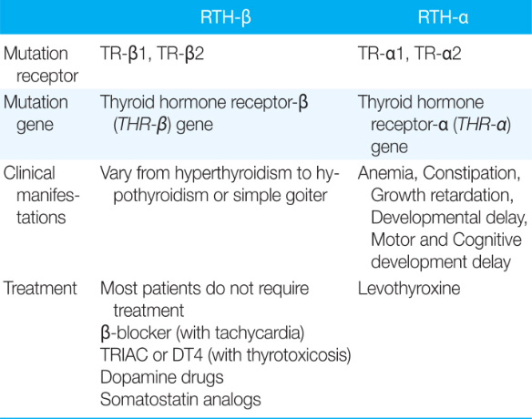
The treatment mainly aims to maintain a balance between the overstimulation of the tissues expressing TRβ in patients with RTH [5]. Currently, no definitive therapy is available to correct the molecular defect that causes RTH syndrome, and most patients do not require treatment [5,9]. Patients with tachycardia and palpitations at rest may benefit from β-blocker therapy. If thyroid toxicity symptoms are present, triiodothyroacetic acid (TRIAC) or dextrothyroxine (DT4) can be administered to treat thyrotoxicosis. This compound, through a feedback mechanism, reduces TSH secretion and decreases circulating T4 levels. TRIAC given at a dose of 1.4–2.8 mg/day divided into two or three administrations is effective in both pediatric and adult patients with RTH [6]. The use of dopamine drugs and somatostatin analogs has limited success because TSH secretion quickly escapes the inhibitory effects of both drugs, as T4 reduction strongly stimulates TSH secretion (Table 1) [4,9].
RTH CAUSED BY THRA MUTATIONS (RTH-α)
RTH-α is characterized by tissue-dependent hypothyroidism findings, and > 40 THRA mutations have been recorded [12]. Most mutations selectively involve the c-terminal domain of TRα, generating severe loss of function and other mutation types associated with a variable-form function of TRα1 [13]. In these cases, the phenotype is characterized by manifestations of hypothyroidism that affects gastrointestinal, neurological, skeletal, and myocardial functions. Clinical characteristics associated with hypothyroidism (e.g., macrocephaly, hypertelorism, flattened nasal bridge, and a broad face) became more evident with time, together with poor linear growth, resulting in a dysmorphic appearance. Patients with RTH-α have normal hormonal response in the pituitary hypothalamic axis and liver. The severity of the clinical phenotype appears to be associated with the mutation location and type in THRA. When heterozygous mutations in this gene are present, abnormalities include anemia, constriction, and delayed growth and development [9]. The A263V mutation of TRα1 showed partial loss of function, with higher T3 levels. TRα2 has no mutation-related phenotypes identified; however, some unusual clinical characteristics, such as micrognasia, clavicular agenesis, hyperparathyroidism, and chronic diarrhea, were reported in an adult woman with a different mutation (N359Y), common for both TRα1 and TRα2 [14]. In RTH-α, serum T3 levels can be high or normal, T4 and rT3 levels are in the normal range or low, and TSH levels are normal or slightly high [3].
Levothyroxine therapy generates the normalization of T4 levels, with additional increase in T3 and suppressed TSH levels, suggesting conserved negative feedback of TH over TSH secretion (Table 1) [3].
CONCLUSION
RTH is a group of genetic-related syndromes that result from different mutations, either in the genes they encode for receptors. These syndromes are characterized by decreased tissue sensitivity to THs, generating various clinical manifestations. Thus, clinical changes of resistance to TH must be recognized and differentiated, and an approach to the practice of personalized medicine through an interdisciplinary approach is needed.