open access
메뉴 E-ISSN : 2671-6771


Infantile nystagmus syndrome (INS) is a genetically heterogeneous disorder. To date, more than 100 genes have been reported to cause INS and there is significant overlap in phenotypic characteristics. The most common form of X-linked INS is attributed to FRMD7 at Xq26. Recent advances in molecular genetics have facilitated the identification of pathogenic variants of FRMD7 and the investigation for underlying mechanisms of FRMD7-associated INS. This review summarizes genetic and clinical features of FRMD7-associated INS, and introduces updates on the pathogenesis of FRMD7 mutation.
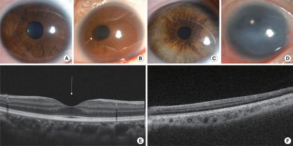
Infantile nystagmus syndrome (INS), formerly called congenital nystagmus is characterized by rhythmic involuntary oscillations of the eyes that are present at birth or during infancy [1]. It can be associated with afferent visual system disorders such as ocular albinism, anterior segment dysgenesis, and foveal hypoplasia (Fig. 1). On the other hand, idiopathic INS arises independently of any other visual or neurological disorders. This has led to speculation that idiopathic INS may be caused by abnormal development of the ocular motor system itself rather than disorders of the afferent visual pathway [2,3].
The inheritance patterns of idiopathic INS are heterogeneous and have been reported as autosomal dominant, autosomal recessive, or X-linked trait [4-6]. However, the most common form of inheritance is X-linked, which can be dominant or recessive. Three loci have been identified at Xp11.4-p11.3, Xp22, and Xq26-q27, but approximately 50% of idiopathic INS families have been linked to Xq26-q27. After Tarpey et al. first identified pathogenic mutations in FRMD7 (MIM#300628) at Xq26, over 90 different mutations have been reported in patients with idiopathic INS [7].
This review summarizes genetic and clinical features of FRMD7-associated INS, and introduces updates on the pathogenesis of FRMD7 mutation.
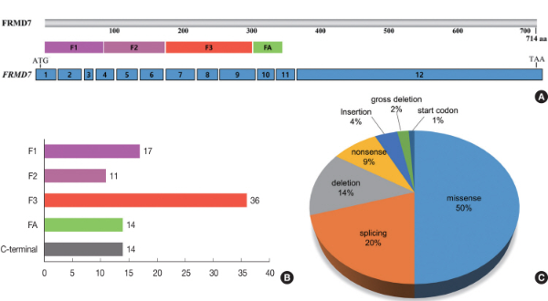
The FRMD7 gene consists of 12 exons and encodes a 714-residue polypeptide [6]. It contains a conserved N-terminal FERM domain (amino acids 2-282) and FERM-adjacent (FA) domain (amino acids 288-336), whereas the C-terminal region has no significant homology to other proteins (Fig. 2A). The FERM domain has 3 lobed “cloverleaf” structures: F1 (lobe A), F2 (lobe B), and F3 (lobe C). These are plasma membrane-cytoskeleton coupling proteins which bind to actin or other cytoskeleton components. The FA domain contains conserved motifs that are potential substrates for kinases, suggesting its regulatory effect in FRMD7 protein. In situ hybridization experiments in human embryonic brain showed FRMD7 expression in neuronal tissues involved in the vestibulo-ocular reflex and optokinetic reflex such as the developing neural retina, optic stalk, otic vesicle, vestibulocochlear nerve, vestibular nucleus, and cerebellum [7-9].
The FRMD7 protein is highly co-localized with the actin of primary neurites in differentiating Neuro2A cells, which promotes elongation of axons and dendrites [10]. Knockdown of FRMD7 protein causes a reduction in average neurite length [11]. Several studies have proposed a mechanism of FRMD7 regulation for neuronal cytoskeletal dynamics. The FRMD7 protein shares close amino acid sequence homology with two other FERM domain containing proteins: FARP1 and FARP2 [6]. They are involved in neurite outgrowth and branching through activating Rho GTPase signaling. Rho GTPases are key regulators of the actin cytoskeleton in eukaryonic cells and mediate morphological changes during neuronal development and plasticity. It was found that wild-type human FRMD7 activated Rac1 signaling by interacting with RhoGDIα, the main regulator of Rho GTPase, while mutant FRMD7 failed to interact with RhoGDIα and to activate Rac1 signaling [12]. Recent studies also demonstrated that FRMD7 regulates the expression of Rac1 in stable SHSY-5Y cells [13], and mutant FRMD7 significantly influences the expression of Rac1, Cdc42, and RhoA during the induction period of human fibroblasts-reprogrammed neurons [14]. Thus, it is possible that FRMD7 is involved in the regulation of neuronal cytoskeletal dynamics through Rho GTPase signaling at the growth cone. Alternatively, the interaction between FRMD7 and calcium/calmodulin-dependent serine protein kinase (CASK) may promote the membrane extension during neurite outgrowth since the function of CASK is to link the plasma membrane to the actin cytoskeleton [15]. Furthermore, FRMD7 is specially localized in starburst cells of the mouse retina and the directional selective (DS) inhibitory input from starburst cells to DS ganglion cells is lost in FRMD7 mutant mice [16]. A recent study found FRMD7 to directly interact with the loop between transmembrane domains 3 and 4 of GABRA2, a type A gamma-aminobutyric acid (GABA) receptor subunit, and colocalization of FRMD7 and GABRA2 was found in the mouse retina [17]. Thus, FRMD7 mutations perturb the interaction between FRMD7 and GABRA2, which may impair GABA inhibitory inputs from starburst cells to DS ganglion cells, eventually leading to the loss of optokinetic reflex that can be seen in INS patients. All of these findings support that nystagmus by FRMD7 mutations may result from defective axogenesis, dendritogenesis, and neuronal guidance in the areas of the brain which control eye movements.
To date, over 90 different mutations within FRMD7 have been reported (Supplementary Table S1) [18,19]. Approximately 84% of mutations concentrates heavily within the N-terminal FERM and FA domain without any consistent hot spots (Fig. 2B). Most have been identified in single case with idiopathic INS, but some mutations including c.41delAGA (p.K14del), c.70G > A (p.G24R), c.875T > C (p.L292P), c.910C > T (p.R303X), and c.1003C > T (p.R335X) have been detected in different racial groups. Especially, c.875T > C (p.L292P) accounted for more than 50% of Korean patients carrying FRMD7 mutations [19]. All patients with c.875T > C came from the same restricted region (Gyeongsangnam-do) of Korea, and shared two single-nucleotide polymorphisms (rs6637934, rs5977623) of exon 12 within FRMD7, suggesting that c.875T > C might have arisen from the founder effect in the Korean population with idiopathic INS.
A half of the mutations are missense which may destabilize the overall structure of FRMD7 protein, while the other half are predicted to cause gross defects at the protein level due to nonsense mutations, frameshift by small deletion or insertion, aberrant splicing, and large intragenic deletion (Fig. 2C). Among the 12 exons, exon 9 represents the most common mutation-rich exon (23%), followed by exon 12 (12%) and exon 8 (11%).
Incomplete penetrance was observed in female carriers, ranged from 30 to 100% [5,20-23]. This phenomenon has been explained by skewed X-inactivation and interactions with disease-modifying genes or environmental factors. Although skewed X-inactivation has consistently been suggested as a mechanism that may influence the penetrance of X-linked disorders in females, some studies have revealed that there was no clear causal link between X-inactivation pattern and phenotype in INS families with FRMD7 mutation [20,21]. Furthermore, affected females showed random X-inactivation, reflecting a tissue mosaicism [22]. Different methylation patterns for the X chromosome were also found between female carriers, implying that a molecular basis for variable methylation might not be involved in the dissimilar penetrance [21]. Further investigations of X-inactivation status of FRMD7 may help understand the incomplete pattern of inheritance.
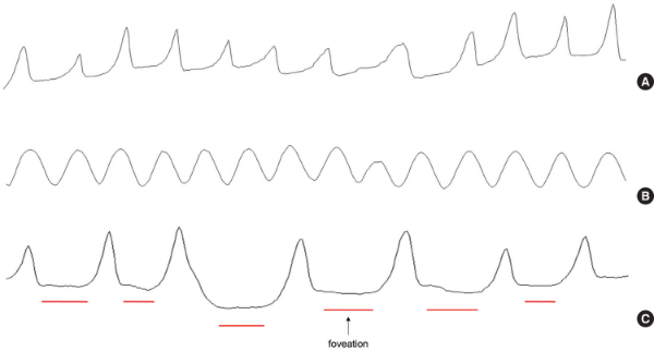
The clinical features of FRMD7-associated INS are not much different from those of non-FRMD7 INS. The nystagmus is present at birth or during infancy, and usually manifests as horizontal conjugate oscillations, while vertical nystagmus is not typical for FRMD7-associated INS [1,4]. The direction of the nystagmus changes with eccentric gaze (right-beating on right gaze and left-beating on left gaze) or alternates periodically with time (periodic alternating nystagmus) [8,19]. The nystagmus is often accentuated by anxiety, attention, and attempts to fixate an object, while attenuated with eyelid closure or on convergence. The nystagmus waveform can be pendular or jerk with increasing exponential slow phases (Fig. 3A and 3B), but a pendular waveform is more common in adults with FRMD7 mutations.
The nystagmus decreases when the eyes are moved into a particular position within the orbit, called the null point or zone [1]. Some individuals with INS tend to turn their head close to the null point or zone, resulting in abnormal head posture (AHP). The presence of AHP often leads parents to bring a child for medical evaluation and treatment.
Despite continuous eye oscillations, individuals with FRMD7-associated INS show relatively good visual acuity and no oscillopsia due to the presence of foveation period which the eye velocity is at or near zero (Fig. 3C). During this brief period, the image of the target is relatively stationary in the foveal area, leading to good visual acuity without oscillopsia [1]. Furthermore, FRMD7-associated INS is not accompanied with afferent visual system disorders causing reduced visual acuity such as foveal anomaly or retina dystrophy. Although previous studies have revealed morphological changes of retina and optic nerve such as decreased peripapillary retinal nerve fiber layer and shallow foveal pit and optic nerve head, these changes may be subclinical [9,18,19]. However, some individuals may complain of oscillopsia when the nystagmus is pronounced or the individual is tired.
FRMD7 is a major disease-causing gene of idiopathic INS. Although the molecular pathogenesis of FRMD7 is still unclear, it is thought that FRMD7 may participate in neuronal development in the areas of the brain controlling ocular motor and gaze stability. Further functional investigation and mutant analysis are needed to reveal the pathogenic mechanisms of FRMD7-associated INS.