
open access
메뉴 E-ISSN : 2671-6771


Pseudogenes are genomic regions that contain gene-like sequences that have a high similarity to the known genes but are nonfunctional. They are categorized into processed, unprocessed, and unitary pseudogenes. Unprocessed pseudogenes generated by duplications can be problematic in sequencing approaches in molecular diagnostics. We discuss the risk of misdiagnosis when investigating genes with pseudogenes of high homology, and describe a method for identifying these small and annoying differences between parent genes and pseudogenes, including parent gene-specific assay design.
Pseudogenes (Ψ) are genomic regions that contain gene-like sequences that have high similarity to the known genes (parent genes, i.e., their functional paralogs) but are non-functional. Therefore, pseudogenes have been considered as ‘junk DNA’ [1-6]. However, recent discoveries unveil this junk DNA’s biological meanings, such as a regulatory role in cancer and developmental processes, and a genetic repository role to store and expand genetic information [7-14]. Explorations and annotations of pseudogenes can give an insight into their genetic evolution, diversification, and functionalization [15-17].
Pseudogenes can be introduced into our genome through several mechanisms such as spontaneous mutation that results in ‘unitary pseudogene’ [18], duplication that constitutes ‘unprocessed pseudogene’ [19], and, retrotransposition of processed mRNAs that produces ‘processed pseudogene’ [20]. Among them, unprocessed pseudogene maintaining its exonic and intronic structure could be problematic in molecular diagnostics because the genetic regions of pseudogenes and parent genes can be co-amplified, co-enriched, and co-aligned to the regions of interest. The mutation rate of pseudogenes is higher than that of the parent genes because they are not often under selection pressure. Therefore, mutations of pseudogenes can misguide the diagnostic approaches to the genetic disorders. Excluding out these false positive mutations with tiny differences (‘nuances’) in their flanking regions is challenging, and, sometimes, missed for geneticists, and, unfortunately, can be reported as a pathogenic variation to clinicians (‘nuisances’).
Here, we discuss the risk of misdiagnosis when investigating genes with pseudogene counterparts of high homology, and we describe the method of identifying these small and annoying differences between parent genes and pseudogenes, including parent gene-specific assay design.
According to the current GENCODE release (version 41, 01. 2022.) that aims to annotate all genetic features at genome-wide level, human genome contains total 61,852 genes including 19,370 protein-coding genes (31%), 19,095 long non-coding RNA genes, 7,566 small non-coding RNA genes, and 14,736 pseudogenes (24%) [21]. Among pseudogenes, processed pseudogenes (10,662, 72%) comprise over two-thirds.
Pseudogenes could be identified by several characteristics such as the absence of introns, truncating mutations, and the absence of transcription. Although most pseudogenes are non-functioning, increasing numbers of pseudogenes have been demonstrated to play some biological roles.
Processed pseudogenes rarely have regulatory elements, thus lack transcriptional activities. PGK2 (chr6:49,785,660-49,787, 285) is a processed pseudogene of phosphoglycerate kinase 1 (PGK1, chrX:77,910,739-78,129,295) via retrotransposition, but, it is expressed specifically in testis, encoding a functional phosphoglycerate kinase that catalyzes the reversible conversion of 1,3-bisphophoglycerate to 3-phophoglycerate, during spermatogenesis [22]. Some processed pseudogenes have intact open reading frames to be able to encode proteins, and play a biological role [23-26].
Unprocessed pseudogenes that arise via duplication preserve regulatory elements and intronic structures, but they are usually non-functional by disruption like truncation mutations. NOTCH2 gene (chr1:119,911,553,120,100,779) has 4 identical paralogs (NOTCH2NLA, NOTCH2NLB, NOTCH2NLC, and NOTCH2NLR) via segmental duplications. NOTCH2NLR is likely to be non-functional, but other 3 NOTCH2NLs are expressed throughout corticogenesis [27]. Interestingly, NOTCH2NLC gene (chr1:149,390,621-149,471,833) is expressed significantly with age [28]. Recently, a heterozygous trinucleotide repeat expansion in the 5’-untranslated regions of the NOTCH2NLC gene was reported to be a causative mutation of neuronal intranuclear inclusion disease (NIID) [29, 30].
DNA sequencing by Sanger method or recent next-generation sequencing (NGS) technology is a crucial step for genetic diagnosis. This strategy can be hampered when there are homology regions with target regions of interest, mostly those are pseudogenes. Mutation data can be erroneous, especially when we analyze target genes with highly homologous unprocessed pseudogene counterparts because segmental duplications can be indistinguishable from their parent region if a laboratory is using short-read sequencing regardless of clinical or research settings.
Autosomal dominant polycystic kidney disease (ADPKD) is a common hereditary kidney disease caused by mutations in PKD1 and PKD2. PKD1 has 6 homologous pseudogenes (PK-D1P1-P6) located between 13 and 16 Mb distant to PKD1 via intrachromosomal duplication. Besides high GC contents and absence of hot spots in PKD1 gene. high sequence homology (98-99%) of pseudogenes complicate the molecular diagnosis of ADPKD [31].
Gaucher disease is an autosomal recessive lysosomal storage disorder caused by mutations of the GBA gene, encoding the lysosomal enzyme acid β-glucosidase. Heterozygous GBA mu-tations are the main genetic risk factor for late-onset Parkinson disease. GBA also has a highly homologous (96-98%) pseudo-gene (GBAP1) located approximately 16 kb distant to GBA [32]. Thus, detecting a pathogenic variant in GBA is challenging with short-read sequencing because of alignment issues [33].
Filamin myopathy is a neuromuscular disorder caused by mutations of FLNC gene, encoding filamin C, muscle-specific filamin isoform cross-linking actin. FLNC has a highly homologous pseudogene (pseFLNC) located 53.6 kb distant to FLNC, which is 98% homologous to exons 46, 47 and 48 of the parent gene [34]. Genetic studies for FLNC had been erroneous for years until Odgerel et al. [35] discerned the misidentification of FLNC mutations and suggested an optimized strategy [36].
A recent study revealed recurrent GNAQ mutation encoding T96S in natural-killer/T cell lymphoma using NGS technologies [37]. However, mutations in this study was revealed to be misaligned to GNAQ instead of GNAQP1, a processed pseudogene of GNAQ [38]. When analyzing somatic mutations using fragmented DNAs, even processed pseudogenes can be problematic.
In the era of NGS technologies, pseudogenes can be still troublesome because short-read sequencing is more susceptible to mis-aligning of homologous sequences than conventional Sanger sequencing. If sequence reads of 150–250 bp containing a pseudogene-derived mutation are mapped to the parent gene, it will generate a false positive result. If sequence reads containing the parent gene-derived variant are mapped to the pseudogene, it will produce a false negative result.
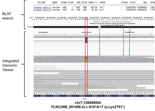
When we devise a diagnostic assay to find a sequence variation in genetic regions of interest with highly homologous pseudogenes, we should confirm that the only desired genetic regions are amplified/enriched, aligned, and analyzed. The first step to do this is to identify small differences in sequence between parent gene and pseudogenes. The BLAST-Like Alignment Tool (BLAT) could be a good starting point to identify small differences [39]. BLAT is typically used to search similar sequence within the same species. Therefore, it is convenient for the identification of discrepant sites between two highly homologous genetic regions.
For example, Fig. 1 demonstrates how to identify the differences between the parent gene (FLNC) and pseudogene (pse-FLNC) in the region of interest. We can annotate the discrepant sequence between parent gene and pseudogene upon the patient’s sequencing reads to discriminate whether a significant variation is from parent gene or pseudogene.
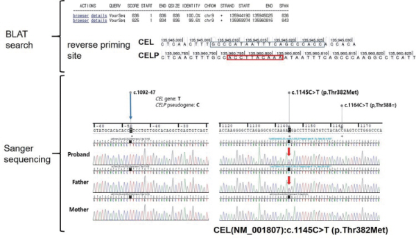
When we cannot discriminate the significant variations from pseudogene-specific alterations in sequencing analysis, we need to find the other orthogonal methods, i.e., parent gene-specific assay. Mostly, we can adopt allele-specific amplification and subsequent Sanger sequencing that can achieve longer sequencing results than NGS. However, sometimes, we need additional separation or enrichment methods, such as cloning. We should manually design all primers and longrange PCR when confirming variants in regions with high homology and devising a new diagnostic assay for an already known gene with highly homologous pseudogenes. Fig. 2 shows the example of parent gene (CEL)-specific assay design and its application in Sanger sequencing. Using the parent gene-specific primer, we can exclude out pseudogene-derived sequence variations.
Even in the era of NGS, specific attention and considerations to discriminate pseudogenes from parent genes are needed. Investigators and clinicians should be aware of the possibility of false positive and/or false-negative results due to highly homologous pseudogenes. Laboratorians and researchers should be prepared for identifying small differences between parent gene- and pseudogene-derived sequences, and designing the parent gene-specific assays.
