
open access
메뉴 E-ISSN : 2671-6771


Background: Arteriovenous malformations (AVMs) are rare diseases comprising abnormally dilated arteries and veins with an absence of a capillary network. Since these diseases are intractable after diagnosis, various treatment strategies have been examined, with continuous efforts to identify target genes. Here, we report relevant new target genes selected via gene microarray. Methods: Endothelial cells were isolated from samples collected from three patients with AVM and three healthy individuals, followed by microarray analysis. Additionally, quantitative PCR was performed to select genes highly relevant to AVM. Results: In the vascular endothelial cells derived from the tissues of patients with AVM, the expression of ANGPT1, ANGPT2, DLL4, IL6, NRG1, TGFBR1, and VEGFA was typically higher compared to those derived from normal tissues. Conclusion: Seven candidate genes were selected to analyze the pathophysiological mechanism of AVM. These results may aid in future directions of diagnosis and treatment.
Among vascular malformations, arteriovenous malformation (AVM) is one of the rare diseases in which the high-pressure blood flow of the artery is directly connected to the draining vein without an intervening capillary network [1-3]. Currently, it is difficult to treat extracranial AVM after diagnosis. To understand the molecular process underlying this intractable rare disease, several studies have examined the basic vascular architecture and differences in the expression of AVM-related biomolecules or angiogenesis-related genes [1,2,4-14]. Expressed genes are being continuously analyzed using the DNA microarray technique based on the results of other studies. Furthermore, treatment strategies are being developed via functional analysis of differentially expressed target genes [9-11]. Accordingly, we report the overexpression of new target genes in extracranial AVM using microarray.
This study was approved by the Institutional Review Board of Kyungpook National University Hospital (approval number: KNUH 2020-03-078-001) and was performed in accordance with the Helsinki Declaration. Sampling was performed after obtaining consent from three patients with extracranial AVM and three healthy individuals. Using paraffin-embedded tissue of arteriovenous vascular malformation, hematoxylin and eosin (H&E), trichrome, elastin, and immunocytochemical staining methods were performed to observe histological morphology.
In the case of vascular endothelial cells, after obtaining patient consent, vascular endothelial tissues to be discarded were obtained after resection. We then cut them with scissors after sterilization and placed the tissue sections in Dispase Ⅱ (17105-041, Gibco), followed by incubation at 4°C for 12 to 24 hours. After soaking the tissue, the epidermis and dermis were separated. Thereafter, the dermal layer was collected in 10 ml of HEPES BSS (14170-112, Invitrogen) and submerged, followed by the removal of the supernatant. A total of 5 mL of collagenase Type Ⅰ was added to the supernatant followed by incubation in a water bath. Subsequently, 10 mL of EMB-2 media (cc-3156, Lonza) was added and the contents were passed through a nylon filter with a pore size of 70 μm. After separating using a centrifuge (2,000 rpm, 3-5 minutes), cells were grown in the EMB-2 media (cc-3156, Lonza). During subculture, vascular endothelial cells were isolated while proliferating the cells using the CD31 Microbead kit (midiMacs starting kit set, 130-042-501).
Molecular biological analysis was performed to confirm the isolation of the vascular endothelial cells. First, RT-PCR found that CD31, a marker of vascular endothelial cells, was strongly expressed in the isolated vestibular endothelial cells when compared with that in the control group. Furthermore, pure vascular endothelial cells were isolated, as demonstrated by the morphology in the cell images. Endothelial cells were extracted from AVM region and normal vessels, and microarray hybridization was performed. Genes related to AVM, such as those participating in angiogenesis and cell proliferation with large fold changes were selected, resulting in a selection of 12 genes with high relevance. After the isolation and culture of vascular endothelial cells derived from patients with AVM patients, PCR amplification products were monitored in real-time. To this end, real-time PCR was performed because accurate quantification of exponential amplification ability is possible, contamination risk is low, and electrophoresis is not required, enabling a rapid and simple interpretation, to verify the expression pattern of target genes that are differently expressed in vascular endothelial cells. A PCR mix was prepared with 7 μl of SYBR Green Master mix (ABI 4367659), 1 μl each of forward primer (10 μM) and reverse primer (10 μM), and 1 μl of 300 ng/μl of cDNA, which was applied to PCR using a real-time PCR ABI 7500. The results were analyzed using Ct values and standard curves.
To identify rarely expressed genes and to select target genes, endothelial cells with proliferative arteriovenous malformation were cultured, followed by RNA isolation. Probe synthesis, hybridization, detection, and scanning of total RNA (5 μg) were performed according to the manufacturer’s instructions (Affymetrix; USA). First, recombinant complementary DNA (cDNA) was synthesized using total RNA. After the cleaning process, double-stranded recombinant cDNA was synthesized. In vitro transcription was performed using the recombinant cDNA as a template and biotin-bound CTP and UTP. After the cleaning process, 10–15 mg of biotin-bound recombinant RNA was fragmented using a fragmentation buffer (Affymetrix, USA). Thereafter, the recombinant RNA was hybridized to the HuGene-1-0-st chip using the standard experimental method (Affymetrix), and then the unbound probe was washed using the GeneChip Fluidics station 450. After washing, the chip was stained with streptavidin-phycoerythrin, and the signal strength was analyzed using the GeneChip scanner 3,000 of Affymetrix and GCOS software to obtain data. Gene expression patterns (Affymetrix microarray data) were analyzed using the Expression console version 1.1 (Affymetrix, USA). Absolute analysis metrics were determined using the Affymetrix Expression Console version 1.1. Change, change p-value, and signal log ratio (fold change) values were obtained by comparison between experimental groups. To reduce the deviation of each value, the number of each experiment was increased to average the fluorescence intensity. RMA-stretch was used for normalization. From the scanning results, genes with up-regulated or down-regulated expression by 1.68-fold or higher were selected. Additional analysis was performed using the upstream regulator analysis in the ingenuity pathway analysis (Ingenuity Systems, USA) methods.
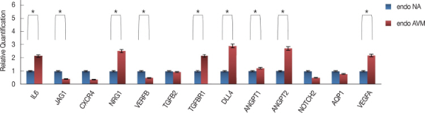
The expression patterns of vascular endothelial cells derived from normal tissues and vascular endothelial cells obtained from tissues of patients with AVM patient tissues analyzed by real-time PCR showed increased expression of ANGPT1, ANGPT2, DLL4, IL6, NRG1, TGFBR1, and VEGFA in vascular endothelial cells of patients with AVM compared to those in normal tissue. Similar or decreased expression of AQP1, CXCR4, JAG1, NOTCH2, TGFB2, and VEGFB genes was observed in the vascular endothelial cells of patients with AVM compared to that in normal tissues. (Table 1, Fig. 1).
| Symbol | Fold change |
Gene | |
|---|---|---|---|
| 7982854 | DLL4 | 0.207 | Delta-like 4 |
| 7955290 | ANGPT1 | 2.738 | Angiopoietin 1 |
| 8145766 | NRG1 | 0.533 | Neuregulin-1 |
| 8119898 | VEGFA | 5.301 | vascular endothelial growing factor-a |
| 8112139 | IL6 | 9.608 | Interleukin-6 |
| 8149071 | ANGPT2 | 0.184 | Angiopoietin 2 |
| 8156826 | TGFBR1 | 1.248 | Transforming growth factor β-receptor 1 |
AVM is one of the rare congenital diseases with common symptoms such as cutaneous stains, local warmth, palpable thrill, and bruit. In later stages, progression to ischemic conditions or indolent ulceration, which can lead to intractable pain, intermittent bleeding, and even congestive heart failure. Treatment typically involves blocking the feeding vessel via embolization. Sclerotherapy, surgical resection, or an appropriate combination of these treatments are also used. In particular, for surgical resection, the risk is low when performed after embolization and 24 to 72 hours before surgery. The success rate is the highest in the case of well-localized AVMs in stages I or II. The best way to determine the resection margin is to clinically examine the bleeding pattern of the wound edges. However, it has a high recurrence rate and may require repeated several surgical treatments. Hence, etiological target genes that can be used to simultaneously diagnose and treat the disease are being continuously examined. In this study, we aimed to report new genes related to AVM by analyzing target genes through microarray. Vascular endothelial cells from patients with AVM and healthy individuals were collected and compared. A dozen genes were found via the selection and analysis of genes associated with both angiogenesis and cell proliferation, which are most relevant processes associated with AVM. Real-time qPCR of such genes showed that the increase in protein levels in the AVM group was statistically significant when compared to those in the control group. A total of seven genes including ANGPT1, ANGPT2, DLL4, IL6, NRG1, TGFBR1, and VEGFA were found to be highly expressed. DLL4, TGFB2, and angiopoietin play an important role in maintaining vascular homeostasis related to angiogenesis and are involved in various processes such as induction of shear stress, hypoxia, and inflammation, which are consistent with AVM [12]. IL6 affects angiogenesis via the PI3K/Akt pathway [13]. In addition, NRG1 encoding neuregulin 1 is used as a treatment for myocardial angiogenesis [14]. In general, the genes that were overexpressed and selected were relevant to angiogenesis, which was also consistent with the clinical characteristics of AVM. Besides, the microarray analysis did not demonstrate increased expression of genes related to other pathways and angiogenesis in AVM in this study. A treatment strategy targeting these seven genes for AVM should be considered. We will continue follow-up studies on drugs or target gene regulators. In conclusion, Seven genes, ANGPT1, ANGPT2, DLL4, IL6, NRG1, TGFBR1, and VEGFA, were selected to analyze the pathophysiological mechanism of AVM. These results would provide the foundation for the development of specific drugs or new treatment methods for patients in the future.
INSTITUTIONAL REVIEW BOARD STATEMENT This study was approved by the Institutional Review Board of Kyungpook National University Hospital (approval number: KNUH 2020-03-078-001) and was performed in accordance with the Helsinki Declaration.
INFORMED CONSENT STATEMENT Informed consent was obtained from all the subjects involved in the study.
