open access
메뉴 E-ISSN : 2671-6771


β-ureidopropionase (β-UP) is an enzyme that catalyzes the final step in the pyrimidine degradation pathway, which converts β-ureidopropionate and β-ureidoisobutyrate into β-alanine and β-aminoisobutyrate, respectively. β-UP deficiency (UPB1D; OMIM # 613161) is an extremely rare autosomal recessive inborn error disease caused by a mutation in the UPB1 gene on chromosome 22q11. To date, approximately 40 cases of UPB1D have been reported worldwide, including one case in Korea. The clinical manifestations of patients with UPB1D are known to be diverse, with a very wide range of manifestations being previously reported; these manifestations include completely asymptomatic, urogenital and colorectal anomalies, or severe neurological involvement, including global developmental delay, microcephaly, early onset psychomotor retardation with dysmorphic features, epilepsy, optic atrophy, retinitis pigmentosa, severely delayed myelination, and cerebellar hypoplasia. Currently, diagnosis of UPB1D is challenging as neurological manifestations, MRI abnormalities, and biochemical analysis for pyrimidine metabolites in the urine, plasma, and cerebrospinal fluid also need to be confirmed by UPB1 gene mutations. Overall, treatment of patients with UPB1D is palliative as there is still no definitive curative treatment available.
β-ureidopropionase (β-UP) deficiency (UPB1D; OMIM # 613161) is an extremely rare autosomal recessive inborn error of pyrimidine metabolism caused by β-UP deficiency that results from a mutation in the UPB1 gene on chromosome 22q11. [1]. To date, approximately 40 cases of UPB1D have been reported worldwide, including one case in Korea. The clinical manifestations of patients with UPB1D are known to be very diverse, with a wide range of manifestations being reported, from asymptomatic to severe neurological involvement [1-3]. Therefore, for the accurate diagnosis of UPB1D, it is important to perform biochemical analysis to assess pyrimidine metabolite levels and to identify mutations in the UPB1 gene by genetic analysis, alongside considering these clinical features; in particular, this is because it is difficult to diagnose UPB1D by assessing symptoms alone. In this paper, the overall biochemical, clinical, and genetic aspects of UPB1D are summarized; therefore, this assessment may increase interest in diagnosing UPB1D patients, and contribute to establishing corresponding treatment methods in the future.
Purine and pyrimidine nucleotides are essential cellular components that are involved in energy transfer and metabolic regulation, alongside DNA and RNA synthesis. Purine and pyrimidine are nitrogenous bases that are major components of different nucleotides in DNA and RNA. Purines are two-carbon nitrogen ring bases, including adenine and guanine, whereas pyrimidines are one-carbon nitrogen ring bases, including cytosine and thymine. Overall, pyrimidine nucleotide metabolism can be divided into three pathways (Fig. 1).
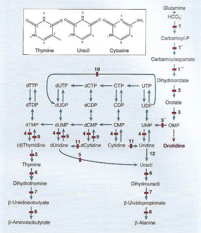
I) The biosynthetic de novo pathway is initiated by the formation of carbamoyl phosphate by cytosolic carbamoyl phosphate synthetase II (CPS II), which differs from mitochondrial CPS I, the latter instead catalyzing the first step of ureagenesis. This formation of carbamoyl phosphate is followed by the synthesis of uridine monophosphate (UMP), then (deoxy)nucleoside triphosphates ((d)NTPs), which are used for RNA and DNA synthesis.
II) The catabolic pathway starts with cytidine monophosphate (CMP), UMP, and thymidine monophosphate (dTMP) and yields β-alanine and β-aminoisobutyrate, which are converted into citric acid cycle intermediates.
III) The salvage pathway, which comprises several kinases, converts the pyrimidine nucleosides cytidine, uridine, and thymidine into their corresponding nucleotides: CMP, UMP, and dTMP, respectively. This pathway also converts several pharmacological anticancer and antiviral nucleoside analogs into their active forms [4].
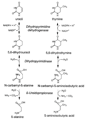
During the pyrimidine metabolic pathway, the pyrimidine bases uracil and thymine are degraded via a three-step pathway, as discussed below (Fig. 2).
I) Dihydropyrimidine dehydrogenase is the initial, ratelimiting, enzyme that catalyzes the reduction of thymine and uracil to 5,6-dihydrothymine and 5,6-dihydrouracil, respectively.
II) The second step of this pathway consists of the hydrolytic ring opening of dihydropyrimidines, catalyzed by dihydropyrimidinase.
III) Finally, the resulting N-carbamyl-β-aminoisobutyric acid and N-carbamyl-β-alanine are converted by β-UP into β-aminoisobutyric acid and β-alanine, respectively, along-side ammonia and CO2 production [5].
The pyrimidine degradation pathway plays an important role in β-alanine and β-aminoisobutyric acid synthesis. β-alanine is a structural analog of γ-aminobutyric acid and glycine, the major inhibitory neurotransmitters in the central nervous system; additionally, β-aminoisobutyric acid has been shown to be a partial agonist of glycine receptors. Therefore, pyrimidine nucleotides are essential for numerous biological processes, such as the synthesis of RNA, DNA, phospholipids, and glycogen, alongside the sialylation and glycosylation of proteins. Further, as pyrimidines play an important role in regulating the central nervous system, changes in pyrimidine metabolism can lead to corresponding abnormal neural activity [5-8].
In 1998, the first reported patient with UPB1D was a 17-month-old girl with hypotonia, dystonia, scoliosis, and severe developmental delay [9]. Enzymatic defects in this patient were later confirmed by enzyme analysis of the liver [10]. Later, in 2002, four patients were reported to have been identified through a urine neonatal screening program; in 2004, three additional patients were identified using high-performance liquid chromatography (HPLC)-tandem mass spectrometry [11,12].
To date, approximately 40 cases of UPB1D have been reported worldwide, including seven cases that have been recently reported in China [4]; the clinical symptoms of these reported UPB1D patients are known to be highly variable. A wide range of clinical symptoms has been reported; these manifestations have been observed to range from completely asymptomatic to urogenital and colorectal anomalies to severe neurological involvement, including global developmental delay, microcephaly, early onset psychomotor retardation with dysmorphic features, epilepsy, optic atrophy, retinitis pigmentosa, severely delayed myelination, and cerebellar hypoplasia [1-3, 11,12]. The clinical features and genetic analysis of UPB1D patients reported to date are summarized in Table 1 [3].
| Patient No. | Origin | Consan-guinity | Sex | Age (yr) | Clinical feature | Genotype | Effect | Location |
|---|---|---|---|---|---|---|---|---|
| 1 | China | − | F | 4 | AS | c.[977G>A]+[977G>A] | p.[R326Q]+[R326Q] | Ex 9 |
| 2 | China | − | M | 0.2 | AS | c.[91G>A+c.977G>A]+[977G>A] | p.[G31S+p.R3260]+[R3260] | Ex 1, Ex 9 |
| 3.1 | China† | − | M | Birth | Transient convulsion | c.[851G>T]+[977G>A] | p.[C284F]+[R3260] | Ex 7, Ex 9 |
| 3.2 | China† | − | F | AS | c.[977G>A]+[977G>A] | p.[R326Q]+]R326Q] | Ex 9 | |
| 4 | China | − | M | 0.4 | MR, polydactyly, MRI-DM | c.[977G>A]+[977G>A] | p.[R326Q]+]R326Q] | Ex 9 |
| 5 | China | − | M | 0.5 | Hypotonia,GR,MR | c.[977G>A]+[977G>A] | p.[R326Q]+]R326Q] | Ex 9 |
| 6 | China | − | M | 0.2 | AS | No DNA available | - | - |
| 7 | China | − | M | 2 | Speech disorder,autism GR MRI-DM | c.[853G>A]+[917-1G>A] | p.[A285T]+splicing | Ex 7, Int 8 |
| 8 | Turkey | + | F | 5.3 | MR, hypotonia, MRI-A, VA, BH, DM,CH | c.[105-2A>G]+[917-1G>A] | splicing | Int1, Int8 |
| 9 | Turkey | + | F | 3 | Seizures, MR,GR, MRI-DM | c.[105-2A>G]+[105-2A>G] | splicing | Int1 |
| 10 | Germany | − | M | 0.9 | Seizures, MC, MR, hypotonia, MRI-A, SH | c.[917-1G>A]+[917-1G>A] | splicing | Int8 |
| 11 | African | − | F | 1 | Seizures | c.[254C>A]+[254C>A] | p.[A85E]+[A85E] | Ex2 |
| 12 | Australia | − | M | 1 | Congenital anomalies of the urogenital and colorectal system | c.[209G>AC]+[105-2A>G] | p.[R70P]+splicing | Ex2, Int1 |
| 13 | Japan | − | F | 0.2 | Seizure (West syndrome), MRI-CD | c.[977G>A]+[977G>A] | p.[R326Q]+]R326Q] | Ex 9 |
| 14.1 | Turkey‡ | + | M | 0.8 | Seizurees, hypotonia | c.[1076C>T]+[1076C>T] | p.[T359M]+[T359M] | Ex10 |
| 14.2 | Turkey‡ | + | F | 30 | AS | c.[1076C>T]+[1076C>T] | p.[T359M]+[T359M] | Ex10 |
| 15.1 | Egypt§ | + | F | neonatus | Seizurees, MC, MRI-CD | c.[105-2A>G]+[105-2A>G] | splicing | Int1 |
| 15.2 | Egypt§ | + | M | neonatus | Seizurees, MC, MRI-CD | No DNA available | - | - |
| 15.3 | Egypt§ | + | M | 27 | AS | c.[105-2A>G]+[105-2A>G] | splicing | Int1 |
| 16 | Egypt | + | M | 0.8 | Seizures, MR, hypotonia, MRI-A | c.[38T>C]+[38T>C] | p[L31S]+[L31S] | Ex1 |
| 17 | Pakistan | + | F | 2 | MC,MR,hypotonia,autism, GR,MRI-A,MRI-CH | c.[792C>A]+[873+1G>A] | p[S264R]+splicing | Ex7,Int7 |
| 18 | China | − | M | 1.1 | MC,MR,GR, MRI-DM | c.[977G>A]+[977G>A] | p.[R326Q]+]R326Q] | Ex 9 |
| 19 | Germany | − | F | 0.9 | Seizures, hypotonia, MRI-A | c.[703G>A]+[917-1G>A] | p.[G235R]+splicing | Ex6,Int8 |
| 20 | China | − | M | 3 | MR,GR | c.[706C>T]+[792C>A] | p.[R236W]+[S264R] | Ex6,Ex7 |
| 21 | Japan | − | M | 3.5 | MR,autism | c.[977G>A]+[977G>A] | p.[R326Q]+]R326Q] | Ex 9 |
| 22 | Japan | − | M | 1 | Motor retardation, hypotonia,MR | c.[811G>A]+[977G>A] | p.[E271K]+R[326Q] | Ex7,Ex9 |
| 23 | Japan | − | M | neonatus | Febrile seizure | c.[977G>A]+[977G>A] | p.[R326Q]+]R326Q] | Ex 9 |
| 24.1 | JapanII | − | F | neonatus | AS, hypermetropia | c.[857T>C]+[977G>A] | p.[I286T]+[R236Q] | Ex7,Ex9 |
| 24.2 | JapanII | − | F | neonatus | AS | c.[857T>C]+[977G>A] | p.[I286T]+[R236Q] | Ex7,Ex9 |
| 25 | Japan | − | M | neonatus | AS | c.[977G>A]+[977G>A] | p.[R326Q]+]R326Q] | Ex 9 |
| 26 | Japan | − | M | neonatus | AS | c.[977G>A]+[977G>A] | p.[R326Q]+]R326Q] | Ex 9 |
| 27 | Japan | − | F | neonatus | AS | c.[977G>A]+[977G>A] | p.[R326Q]+]R326Q] | Ex 9 |
| 28 | Japan | − | M | neonatus | AS | c.[91G>A]+[977G>A] | p.[G31S]+[R326Q] | Ex1,Ex9 |
| 29 | Japan | − | F | neonatus | AS | c.[977G>A]+[977G>A] | p.[R326Q]+]R326Q] | Ex 9 |
| 30 | Japan | − | F | neonatus | AS | c.[977G>A]+[977G>A] | p.[R326Q]+]R326Q] | Ex 9 |
| 31 | Japan | − | F | neonatus | AS | c.[91G>A]+[977G>A] | p.[G31S]+[R326Q] | Ex1,Ex9 |
| 32 | Korea | − | F | neonatus | Seizure, MR,GR,MC,hypotonia | c.[91G>A]+[977G>A] | p.[G31S]+[R326Q] | Ex1,Ex9 |
| 33 | Japan | − | F | neonatus | Hypotonia,MR,GR, MRI-diffuse brain atrophy | c.[976C>T]+[977G>A] | p.[R326W]+[R326Q] | Ex 9 |
A, atrophia cerebri; AS, asymptomatic; BH, brainstem hypoplasia; CD, cortical dysplasia; CH, callosal body hypoplasia; DM, delayed myelination; GR, growth retardation; MC, microcephaly; MR, mental retardation; NA, not available; VH, vermis pypoplasia.
†The same family members (child and father).
‡The same family members (child and mother).
§The same family members (two siblings and one father).
IIThe same family members (twin siblings).
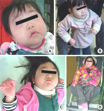
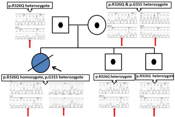
A 3-month-old girl born to unrelated Korean parents visited an outpatient department after experiencing a generalized tonic-clonic seizure without fever. The family history was nonspecific, with a normal vaginal delivery at 41 weeks, a birth weight of 3,140 g (40th percentile), a height of 51 cm (70th percentile), and a head circumference of 32.5 cm (15th percentile). The seizures of the patient were effectively controlled and stopped after phenobarbital treatment. The development of the patient after these seizures was normal; however, seizures recurred at 12 months of age. We performed tests, such as electroencephalography (EEG), brain magnetic resonance imaging (MRI), and tandem mass screening, but no abnormal findings were observed. These seizures could not be controlled despite the use of multiple antiseizure medications, pyridoxine, and a ketogenic diet. Afterwards, the patient exhibited microcephaly, with a head circumference less than the 1st percentile, and global developmental delay. We performed the Korean Bayley Scale of Infant Development-II (K-BSID-II) test at 27 months of age; the corresponding results determined that this patient possessed a mental performance level of a 4-month-old, alongside a motor performance level of a 6-month-old. Additionally, the patient developed generalized status epilepticus at 3-yearsold. Additional diagnostic tests, including a MeCP2 gene test for Rett syndrome, methylation test for Prader-Willi/Angelman syndrome, and muscle biopsy for mitochondrial disease, were all determined to be normal. At age 8, corresponding neurological development deteriorated, with various symptoms including generalized hypotonia, an inability to speak or sit up-right independently; additionally, the patient continued to experience intractable epilepsy (Fig. 3). At that time, we also sent the patient’s DNA sample to the Seattle Children’s Hospital Research Institute for analysis by target panel testing. The UPB1 gene for UPB1D was one of the genes included in this panel, which revealed that she was homozygous for p.R326Q (c.977G >A) and heterozygous for p.G31S (c.91G>A) in UPB1 (confirmed by Sanger sequencing analysis, NM_016327). The corresponding family was subsequently subjected to sequencing analysis, which revealed heterozygosity for p.R326Q in the father and for both p.R326Q and p.G31S in the mother. The two asymptomatic siblings were both heterozygous for p.R326Q (Fig. 4). Biochemical analysis of pyrimidine metabolites could not be performed in Korea; therefore, patient serum, urine, and cerebrospinal fluid (CSF) samples were sent to the Netherlands for analysis. The corresponding results revealed highly elevated N-carbamyl-β-amino acids and moderately elevated levels of dihydrothymine in the CSF and plasma, thereby supporting the biochemical diagnosis of UPB1D (Table 2).
| Urine (umol/mmol creatinine) | Plasma (μM) | Cerebrospinal fluid (μM) | ||||
|---|---|---|---|---|---|---|
| Patient | Reference | Patient | Reference | Patient | Reference | |
| Pyrimidine metabolites | ||||||
| N-carbamyl-B-alanine | 248 | 11.0 ± 9.2 | 6.81 | 0.2 ± 0.3 | 0.90 | 0.1 ± 0.3 |
| N-carbamyl-B-aminoisobutyric acid | 186 | 1.8 ± 2.3 | 25.15 | 0.1 ± 0.2 | 1.60 | 0.01 ± 0.04 |
| Dihydrouracil | 16 | 6.3 ± 5.3 | 0.81 | 1.3 ± 0.8 | 1.60 | 2.1 ± 1.0 |
| Dihydrothymine | 47 | 3.1 ± 2.1 | 3.58 | 0.9 ± 0.3 | 4.30 | 1.1 ± 0.3 |
| Uracil | 4 | 11.8 ± 9.1 | < 0.6 | 0.2 ± 0.4 | 0.20 | 0.1 ± 0.2 |
| Thymine | 1 | 0.5 ± 0.6 | < 0.08 | 0.05 ± 0.03 | < 0.1 | < 0.1 |
The reference values are depicted as mean ± SD.
Physical measurements taken at the age of 8 years and 10 months demonstrated that the patient possessed failure to thrive, global developmental delay, and microcephaly. She had a body weight of 11.2 kg (<1st percentile), height of 103 cm (<1st percentile), and a head circumference of 44.5 cm (<1st percentile). After being diagnosed with UPB1D, the patient was placed on a purine-restricted diet because a pyrimidine-free diet was not possible. Following this dietary restriction, the frequency of seizures and severe seizures, such as status epilepticus, moderately decreased; nonetheless, these seizures were still not effectively controlled. This patient died suddenly in her sleep of unknown cause at the age of 11 years and 5 months [13, 14].
1) Clinical manifestations: The clinical symptoms of UPB1D patients are highly variable, ranging from asymptomatic to severe neurological symptoms. Therefore, it is difficult to make a diagnosis based on clinical symptoms alone. However, if there are UPB1D-associated symptoms, such as global developmental delay, intractable epilepsy, microcephaly, early onset psychomotor retardation with dysmorphic features, optic atrophy, retinitis pigmentosa, severely delayed myelination, cerebellar hypoplasia, or urogenital and colorectal anomalies, for which no cause can be found, a diagnosis of UPB1D must be made. The author proposes to consider this possibility of UPB1D and proceed with further evaluation.
2) Biochemical analysis of pyrimidine bases and metabolites in the body fluids: For the diagnosis of patients with UPB1D, it is particularly important to identify levels of pyrimidine bases, uracil and thymine, and their corresponding degradation products in urine, plasma, and CSF samples. Patients with UPB1D possess normal or slightly elevated uracil and thymine levels, while dihydropyrimidine concentrations are slightly elevated. In contrast, the concentrations of N-carbamyl-β-alanine and N-carbamyl-β-aminoisobutyric acid are strongly elevated compared to those observed in control groups [3, 5, 13]. If it is difficult to clinically identify the pyrimidine degradation products in the urine, plasma, and CSF, it is understood that UPB1D patient screening by the identification of pyrimidine degradation products in the urine is particularly useful for diagnoses [3].
3) β-UP activity in a liver biopsy: In 2004, van Kuilenburg et al. confirmed that in liver biopsies obtained from UPB1D patients, β-UP activity was observed to be normal in controls but indetectable in patients. In contrast, liver biopsies from UPB1D patients exhibited normal activity of dihydropyrimidine dehydrogenase compared to that in controls.
4) Genetic test for UPB1 mutations: To diagnose UPB1D patients, genetic analysis to confirm UPB1 mutation is essential. As of 2019, 18 homozygous or compound heterozygous mutations in UPB1 have been reported in 38 patients (33 families) diagnosed with UPB1D (Table 1).
Treatment of UPB1D patients remains palliative. Additionally, the treatment of UPB1D patients with β-alanine for more than 1.5 years was determined to not lead to any observable clinical improvement [15]. Nonetheless, administration of β-UP for supplementation of β-aminoisobutyric acid and elimination of N-carbamyl-β-alanine and N-carbamyl-β-aminoisobutyric acid is yet to be attempted in patients with UPB1D. Further, a purine-restricted diet may be a potential therapeutic option for treating patients with UPB1D and may reduce the frequency and intensity of seizures [13].
UPB1D is an extremely rare autosomal recessive congenital disorder caused by mutations in UPB1 [1]. Approximately 40 cases of UPB1D have been reported worldwide, including one Korean patient. The corresponding clinical symptoms are understood to be very diverse, with clinical symptoms ranging from completely asymptomatic to urogenital and colorectal malformations to severe neurological involvement, including global developmental delay, microcephaly, refractory epilepsy, delayed myelination, and cerebellar hypoplasia. UPB1D diagnosis is based on clinical symptoms, biochemical analysis of pyrimidine metabolites, and identification of mutations in the UPB1 gene. Currently, screening via the biochemical analysis of pyrimidine metabolites in urine is considered important for identifying these patients. Although the treatment of UPB1D patients remains palliative, it is hoped that a suitable treatment method will be established if more patients are diagnosed and studied in the future.
FINANCIAL DISCLOSURE STATEMENT This review did not receive any specific grant from funding agencies in the public, commercial, or not-for-profit sectors.
We would like to thank the Korean patients and their parents, and also thank Editage ( www.editage.co.kr) for helping with English editing.
An Online Catalog of Human Genes and Genetic Disorders (OMIM®) (2023) Baltimore: University of Johns Hopkins https://www.omim.org/entry/613161?search=UREIDOPROPIONASE&highlight=ureidopropionase, Internet, updated on 2023 Feb 23