
open access
메뉴


Background: Ca2+ signaling plays a vital role in neuronal signaling and altered Ca2+ homeostasis in Parkinson’s disease (PD). Over-expression of αSYN significantly promote the Ca2+-Calmodulin (CaM) activity and subsequent nuclear translocation of nuclear factor of activated T cells (NFAT) transcription factor in dopaminergic neurons of midbrain. However, the exact role of Ca2+-CaM and NFAT in PD pathology is yet to be elucidated. Methods: We designed the CaM-NFAT-oligodeoxynucleotide (ODN), a synthetic short DNA containing complementary sequence for NFAT transcription factor and CaM mRNA. Then, the effect of CaM-NFAT-ODN on 1-methyl-4-phenylpyridinium (MPP+)-mediated neurotoxicity was investigated in mimic PD model in vitro. Results: First, the expression of αSYN and CaM was strongly increased in substantia nigra (SN) of PD and the expression of tyrosine hydroxylase (TH) was strongly increased in control SN. Additionally, the expression of apoptosis marker proteins was strongly increased in SN of PD. Transfection of CaM-NFAT-ODN repressed CaM and pNFAT, the target genes of this ODN in rat embryo primary mesencephalic neurons. It also reduced ERK phosphorylation, a downstream target of these genes. These results demonstrated that CaM-NFAT-ODN operated successfully in rat embryo primary mesencephalic neurons. Transfection of CaM-NFAT-ODN repressed TH reduction, αSYN accumulation, and apoptosis by MPP+-induced neurotoxicity response through Ca2+ signaling and mitogen-activated protein kinases (MAPK) signaling. Conclusion: Synthetic CaM-NFAT-ODN has substantial therapeutic feasibility for the treatment of neurodegenerative diseases.
Parkinson’s disease (PD) is one of the most prevalent neurodegenerative disorders [1]. The pathology of the disease is the selective and progressive loss of the dopaminergic neurons, leading to the formation of Lewy bodies in substantia nigra (SN) pars compacta of midbrain [2]. Lewy bodies are formed by fibrils of alpha-synuclein (αSYN), and this cascade is an important step in the development of PD [3]. Aggregated αSYN regulates dopamine biosynthesis by triggering dopamine transporter (DAT) recruitment of dopamine and reducing the activity or the phosphorylation status of tyrosine hydroxylase (TH), thus leading to neuronal demise [4]. To date, no drugs have been developed to alleviate the loss of dopaminergic neurons in PD and the pathogenesis of PD remains controversial.
Ca2+ homeostasis plays a key role in the degeneration of dopaminergic neurons [5]. Additionally, the increase in cytosolic Ca2+ is dependent on Ca2+ channel activity and αSYN expression, and these two pathogenic factors were reported to act in PD [4]. The degeneration of dopaminergic neurons is a major contributor to the pathogenesis of midbrain disorders [6]. Ca2+ is important for many cellular processes, such as proliferation, growth, differentiation, development, and cell death [7]. Calcium signaling is essential for regulating dopamine concentration evels in neurons [8]. Exposure to Ca2+ overload has been reported to be an important determinant, and pharmacological treatments that inhibits Ca2+ entry reduce neuronal damage in chemical models of PD [9]. Calmodulin (CaM), the best studied Ca2+-binding protein, is abundantly expressed in the brain [10]. CaM contains globular domains at its C- and N-terminus respectively [11]. Each globular domain of CaM has a pair of Ca2+ binding sequences, and after binding Ca2+, a hydrophobic surface is exposed [12]. The expression of CaM is up regulated in response to the increase in intracellular Ca2+ concentration and can thus lead to NFAT (nuclear factor of activated T-cells) dephosphorylation and nuclear translocation [9].
Recent reports have highlighted the important role of the NFAT transcription factor family in neurons involved in the regulation of synaptic plasticity, axonal growth and neuronal survival [13]. The NFAT regulatory domain is highly phosphorylated in the inactive state, covering the nuclear localization sequence and causing the NFAT protein to remain in the cytoplasm [14]. NFAT proteins directly regulate the expression of apoptosis-related genes and with the promotion of inflammatory process [15]. Both apoptosis and inflammation are processes widely involved in the development of PD [7,16].
The synthetic oligodeoxynucleotide (ODN) technique is a gene therapy strategy that DNA or RNA based molecular compounds that are utilized to disrupt gene transcription or translation [17]. Antisense ODN are used for selective impairment of protein synthesis based on the specific sequence of the target RNA [18,19]. Decoy ODN is employed to block transcription factor activity using synthetic double stranded ODN containing DNA binding sites, which works at the pre-transcription level [20,21]. Advances in gene therapy strategies have provided new technologies to inhibit dual-target gene expression based on short RNA and DNA strategies [22,23]. To improve a new therapeutic approach, in this study we used a combination of antisense ODN and decoy ODN to synthesize CaM-NFAT-ODN, which inhibits both CaM and NFAT. Although these ODN have proven beneficial in several disease models, it has not yet been demonstrated whether CaM-NFAT-ODN can attenuate the development of molecular mechanisms of neurotoxicity. Therefore, we investigated the effect of CaM-NFAT-ODN on neuronal cytotoxicity in an in vitro model of Parkinson’s disease.
Synthetic ODNs were commissioned by Macrogen (Seoul, Korea). To generate the ODN of CaM, it was selected through sequential overlapping simulations of the secondary structure using the S-Fold program. The sequence of CaM antisense ODN is 5´-GAA TTC GGT ATC AAG AAC TAG ATA TAC C-3´ and the sequence of NFAT decoy ODN is 5´-GAA TTC TGA CTT TCC TCA TGA AAA CAT GAG GAA AGT AC-3´. The underline is the target site as well as the agreed combining order. These ODNs were structured by annealing and each ODN was mixed with T4 ligase (Takara Bio, Otsu, Japan) to obtain a covalent ligation for ring-type ODN molecules.
A total of eight postmortem brain samples were obtained (Tables 1 and 2); with clinically and neuropathologically diagnosed PD and controls. Brain sections and tissues were provided by the Korea Brain Bank Network (KBBN) operated through the National Brain Bank Project funded by the Ministry of Science and ICT. The project was approved by the Daegu Catholic University Institutional Review Board, DCU IRB (IRB number: CR-20-149).
| KBBN ID | Age | Gender | Final diagnosis | Tissue |
|---|---|---|---|---|
| 07004509 | 50–59 | F | Control | SN |
| 07004529 | 60–69 | M | PD | SN |
F, Female; M, Male; SN, Midbrain with substantia nigra; PD, Parkinson disease.
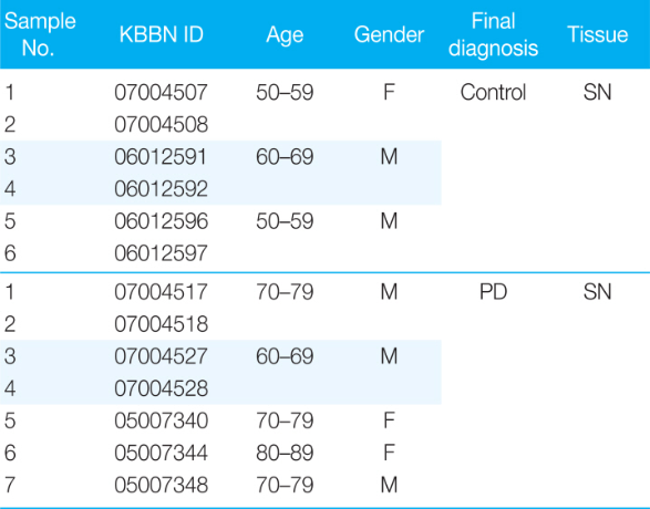
F, Female; M, Male; SN, Midbrain with substantia nigra; PD, Parkinson disease.
Mesencephalic neuron cultures were prepared from the ventral mesencephalic tissues of embryonic day 13–14 rats, as described previously [7,24]. All experimental protocols were approved by the Institutional Animal Care and Use Committee of the Daegu Catholic University Medical Center (EXP-IRB number: DCIAFCR-191112-07-Y) in accordance with criteria out-lined in the Institutional Guidelines for Animal Research. Briefly, dissociated cells were seeded on poly-d-lysine and laminin-coated plates. Cells were cultured in a Dulbecco’s modified Eagle’s medium/Ham’s F-12 medium (Gibco, NY, USA) containing ITS premix (Sigma-Aldrich, St Louis, MO, USA) and 1% penicillin-streptomycin (Gibco).
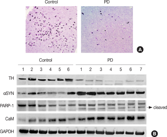
Rat embryo primary mesencephalic neurons were transfected with synthetic ODN using Lipofectamine 2000 (Invitrogen, Carlsbad, CA, USA) according to the manufacturer’s instructions. The transfection efficiency of synthetic ODN was observed using ODN labeled with fluorescein isothiocyanate (FITC) using the Label IT nucleic acid labeling kit (Mirus Bio, Madison, WI, USA). After transfection, the rat embryo primary mesencephalic neurons were cultured in 50 μM of MPP+ for 24 hr.
Nissl bodies in human brain slides were detected by a previously described Nissl staining method [25]. Nissl Staining Solution (Cresyl violet stain solution; Abcam, Cambridge, MA, USA) manufacturer’s instructions. Tissues sections were washed in cold phosphate-buffered saline (PBS) for 15 min and block with universal blocking solution (0.3% TritonTM X-100, 1% BSA, 0.05% Tween 20, 0.1% cold fish gelatin and 0.05% sodium azide in PBS) for 1 hr at room temperature.
The morphology of rat embryo primary mesencephalic neurons was observed using an inverted phase contrast microscope (Olympus CKX41SF, Tokyo, Japan, ×200 or ×400 magnification).
Protein samples were prepared from human brain tissue of the SN (Table 2) and rat embryo primary mesencephalic neuron with a protein extraction buffer (N-PERTM, Thermo Fisher Scientific, Waltham, MA, USA) according to the instruction manual. The protein samples were separated on precast gradient polyacrylamide gels (BoltTM 4%–12% Bis-Tris Plus Gels; Thermo Fisher Scientific) and transferred to nitrocellulose membranes (GE Healthcare, Madison, WI, USA) by using BoltTM Mini Blot Module and Mini Gel Tank (Thermo Fisher Scientific), according to the manufacturer’s recommendations. The membrane blocked with 5% bovine serum albumin was probed with a primary antibody and horseradish peroxidase-conjugated secondary antibody. Following a repeat of the wash step, the membrane was kept in enhanced chemiluminescence detection reagents (Thermo Fisher Scientific). Signal intensity was measured with an image analyzer (ChemiDocTM XRS+; Bio-Rad Laboratories). The protein expression values were normalized to GAPDH and β-actin expression values. The primary antibodies used were anti-TH (Cat no: 13106, Cell Signaling Technology, Danvers, MA, USA), anti-αSYN (Cat no: 2628, Cell Signaling Technology), anti-PARP (Cat no: 9542, Cell Signaling Technology), anti-CaM (Cat no: sc-137079, Santa Cruz Biotechnology, TX, USA), anti-pNFATc3 (Cat no: sc-8405, Santa Cruz Biotechnology), anti-NFATc3 (Cat no: sc-365883, Santa Cruz Biotechnology), anti-pERK (Cat no: 4377, Cell Signaling Technology), anti-ERK (Cat no: 4695, Cell Signaling Technology) and anti-β-actin (Cat no: SAB3500350, Sigma-Aldrich), anti-GAPDH (Cat no: sc-47724, Santa Cruz Biotechnology).
Human brain tissues were deparaffinized in xylene and subjected to citrate antigen retrieval prior to immunohistochemistry [25]. Cells were incubated with primary antibodies for 1 hr at room temperature and they were incubated with the Alexa Flour 488 (excitation/emission = 495/519 nm, green, Invitrogen, CA, USA) and Alexa Flour 594 (excitation/emission= 590/617 nm, red, Invitrogen) for 30 min at room temperature. Cells were counterstained with DAPI (excitation/emission= 330–380 nm/460 nm, ImmunoChemistry, MN, USA). Slides were mounted using ProLong® Gold antifade reagent (Molecular Probes® by Life TechnologiesTM, CA, USA). Immunolabeling was examined using an Eclipse Ti-U and confocal microscope (Nikon, Tokyo, Japan).
Increase in cytosolic Ca2+ is dependent on Ca2+ channel activity and αSYN expression [4]. To determine whether these findings are relevant to PD, we examined human postmortem SN from control versus PD patients. Nissl-stained positive neurons in the SN were significantly reduced in the PD brain compared to human control brain (Table 1 and Fig. 1A). αSYN and CaM expression was strongly increased in SN of PD, and expression of TH, a dopaminergic neuron marker, was strongly increased in control SN (Table 2 and Fig. 1B). Cleaved-PARP-1, an apoptosis marker protein, was also confirmed to be strongly increased in SN of PD (Fig. 1B). These results demonstrated that CaM is related to apoptosis of dopaminergic neurons and PD.
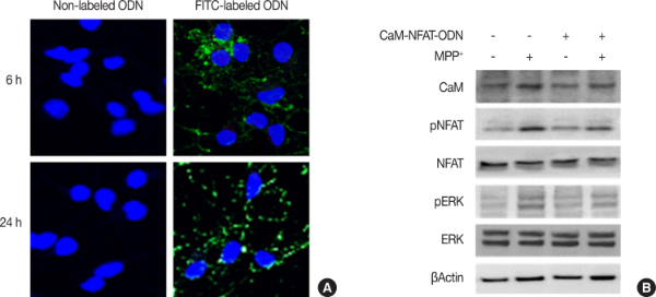
To confirm the efficacy and distribution of the CaM-NFAT-ODN, we transfected rat embryo primary mesencephalic neurons with FITC-labeled ODN. As shown in Fig. 2A, FITC-CaM-NFAT-ODN was successfully transfected and delivered into the nucleus and cytoplasm of rat embryo primary mesencephalic neurons and observed for 24 hr. Moreover, immunoblot analysis showed that the CaM-NFAT-ODN decreased the protein levels of CaM, pNFAT, as well as the key downstream targets of ERK phosphorylation (Fig. 2B).
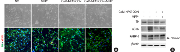
To determine whether Synthetic CaM-NFAT-ODN could regulate the expression of MPP+-induced TH and αSYN in rat embryo primary mesencephalic neurons. The rat embryo primary mesencephalic neurons showed obvious neurites, and the neurons transfected with only CaM-NFAT-ODN were observed similar to normal neurons (Fig. 3A). When rat embryo primary mesencephalic neurons were exposed to the neurotoxin MPP+, neurites were reduced, and neuronal debris increased. Interestingly, it was rescued by CaM-NFAT ODN transfection.
Rat embryo primary mesencephalic neurons were confirmed to be healthy TH-positive neurons with extensive neurites through immunofluorescence staining using a TH antibody. However, exposure to neurotoxin for 24 hr decreased the number of TH-positive neurons and induced accumulation of αSYN. When transfected with CaM-NFAT ODN, the number and morphology of TH-positive neurons were similar to normal neurons. Exposure of MPP+ in CaM-NFAT-ODN transfected neurons appears to protect the cells from loss of TH-positive neurons and accumulation of αSYN. Consistent with immunofluorescence staining, CaM-NFAT-ODN attenuated TH reduction and accumulation of αSYN in rat embryo primary mesencephalic neurons (Fig. 3B). CaM-NFAT-ODN inhibited MPP+-induced cleaved PARP-1, apoptosis marker proteins, in rat embryo primary mesencephalic neurons.
Appearance of PD symptoms is attributed to the loss of dopaminergic neurons in the striatum and substantia nigra [10]. Regarding PD, many CaM-regulated Ca2+ channels seem to be implicated in this pathology [26]. Neuronal cell death may be due to increased Ca2+, which is commonly observed in neurons in various areas of PD brain [5,8]. Additionally, the increase in cytosolic Ca2+ is not due to MPP+-induced oxidative stress but is dependent on Ca2+ channel activity and αSYN expression, and these two pathogenic factors have been reported to act in PD [27]. Our result showed that αSYN and CaM expression was strongly increased in Parkinson’s disease SN, and expression of TH, a dopaminergic neuron marker, was strongly increased in control SN.
A Study has reported on potential neuroprotective agents through attenuation of MPP+ and Ca2+-overload-induced excitotoxicity in dopaminergic neurons [28]. Many reports suggest that Ca2+ is involved in the pathogenesis of PD and that regulation of Ca2+ may be a potential therapeutic target for neuroprotection in PD [29]. For an effective treatment that targets the causes of PD, it is necessary to systemically determine the therapeutic component based on the inhibition of initial molecule mechanism.
We have reported the anti-inflammatory and anti-fibrotic properties of synthetic targeted ODN in chronic diseases [17, 20,21,23]. Recently, we also reported the effects of a selective antagonist of the small-conductivity Ca2+-activated K+ channels antagonist on neuroinflammation and neurotoxicity [7,30]. However, the molecular mechanisms of MPP+-induced neurotoxicity of ODN in PD models have not yet been elucidated.
In this study, we investigated the effects of CaM-NFAT-ODN, which simultaneously regulates the NFAT and CaM, on the neurotoxicity of rat embryo primary mesencephalic neurons.
In PC12 cells, MPP+-induced loss of cell viability was mediated by increased intracellular Ca2+ levels and CaM action [31]. Overexpression of αSYN activates the CaM-NFAT pathway in cell lines and dopaminergic neurons, whereas inhibition of this pathway protects dopaminergic neurons against αSYN-mediated toxicity [32]. The Ca2+-CaM-ERK mitogen-activated protein kinase signaling pathway induces neuronal cell death in rat brain [33]. Our result showed that CaM-NFAT-ODN inhibited ERK phosphorylation, reduction of TH, αSYN expression and apoptosis in MPP+-induced rat embryo primary mesencephalic neurons. Thus, gene therapy targeted to suppress mRNA level of CaM and transcription activity of NFAT simultaneously might provide a new therapeutic strategy to prevent various neurological disorders.
