open access
메뉴


Background: The most prominent pathological features of Parkinson's disease (PD) are diminished substantia nigra (SN), which is part of the output component of the basal ganglia, the severe death of dopaminergic neuronal cell and the accumulation of a synuclein (αSYN). However, the mechanism by which αSYN causes toxicity and contributes to neuronal death remains unclear. Methods: The aim of this study was to investigate the effect of αSYN/STAT oligodeoxynucleotide (ODN), which simultaneously suppresses STAT transcription factors and αSYN mRNA expression in an in vitro Parkinson's disease model. Results: Synthetic αSYN/STAT ODN effectively inhibits 1-Methyl-4-phenylpyridinium (MPP+) induced STAT phosphorylation and αSYN expression. αSYN/STAT ODN attenuated MPP+ to mimic PD model in vitro. MPP+ induced the secretion of TNF-α/IL-6, inhibited cell viability and induced apoptosis while these effects could be rescued by αSYN/STAT ODN. Conclusion: Therefore, synthetic αSYN/STAT ODN has substantial therapeutic feasibility for the treatment of neurodegenerative diseases.
Parkinson’s disease (PD) is a chronic, widespread neurodegenerative disorder characterized by a progressive loss of dopaminergic neurons in the substantia nigra (SN) pars compacta (SNpc) throughout the midbrain [1]. The main pathology of PD is the aggregation of the protein α-synuclein (αSYN) in the cytoplasmic region of dopamine neurons [2].
The Janus Kinase/Signal Transducers and Activators of Transcription (JAK/STAT) is activated by cytokines, interferons, and growth factors [3] and is involved in cell survival, proliferation, angiogenesis, inflammation, and apoptosis [4]. Abnormal activation of JAK/STAT occurs in neuroinflammation and neurodegenerative diseases such as multiple sclerosis, Alzheimer’s disease, and PD [5].
Synthetic oligodeoxynucleotide (ODN) technology is a gene therapy strategy consisting of DNA or RNA-based molecular compounds that disrupt gene transcription or translation [6]. To improve a new therapeutic approach, in this study we used a combination of antisense ODN and decoy ODN to synthesize αSYN/STAT ODN which inhibits both SYN and STAT. Although these ODN have proven beneficial in several disease models, it has not yet been demonstrated whether αSYN/STAT ODN can attenuate the development of molecular mechanisms of neurotoxicity. Therefore, we investigated the effect of αSYN/STAT ODN on neuronal cytotoxicity in an in vitro model of Parkinson’s disease.
Synthetic ODNs were commissioned by Macrogen (Seoul, Korea). The synthetic decoy ODN sequences were used as follows (the target site of the consensus sequence is underlined): STAT decoy ODN: 5´ GAA TTC GTT TCC GGG AAT GAA AAC ATT CCC GGA AAC 3´; αSYN antisense ODN: 5´ GGT ACC CTT CTT CAC CCT TAC C 3´; scrambled (SCR) decoy ODN: 5´ GAA TTC AAT TCA GGG TAC GGC AAA AAA TTG CCG TAC CCT GAA TT 3´. Considering the stability of the decoy ODN strategy, we designed a ring-type structured decoy ODN. These ODNs were annealed for 6 hours while temperature was gradually decreased from 80°C to 25°C. Each ODN was mixed with T4 ligase (Takara Bio, Otsu, Japan) and incubated for 18 hours at 16°C to obtain a covalent ligation for the ring-type decoy ODNs.
A dopaminergic human neuroblastoma cell line SH-SY5Y (America Tissue Culture Collection, CRL-2266; ATCC, Manassas, VA, USA), was cultured in a Dulbecco’s Modi-fied Eagle’s Medium (DMEM) medium (Gibco, Grand Island, NY, USA) containing 10% fetal bovine serum (FBS, Gibco) and 1% Anti-Anti (Gibco). Cell cultures were maintained at 37°C in a humidified atmosphere of 5% CO2. The sources of the following reagents were: 1-Methyl-4-phenylpyridinium ion (MPP+) (Sigma-Aldrich); anti-SYN (Cat no: 2628, Cell Signaling Technology), anti-PARP-1 (Cat no: 9542, Cell Signaling Technology), anti-pSTAT3 (Cat no: 9145, Cell Signaling Technology) and anti-β-actin (Cat no: SAB3500350, Sigma-Aldrich). Immunoblots were detected using an enhanced chemiluminescence reagent (Amer-sham Bioscience, Amersham, UK).
To evaluate the effect of αSYN/STAT ODN on MPP+ stimulated proliferation SH-SY5Y cells were plated in 96-well culture plates at 1×10⁵ cells/ml in culture medium and allowed to attach for 24 hours. Media were discarded and transfect with αSYN/STAT ODN in a new medium, then treat with MPP+ for 24 hours. Cell viability was analyzed using the Cell Counting Kit (CCK-8; Dojindo Laboratories, Kumamoto, Japan) assay according to the manufacturer’s instructions. The absorbance at 450 nm was assessed using a microplate reader (Thermo Fisher Scientific, Waltham, MA, USA).
SH-SY5Y cells were transfected with synthetic ODN using Lipofectamine 2000 (Invitrogen, Carlsbad, CA, USA) according to the manufacturer’s instructions. After transfection, the SH-SY5Y cells were cultured in MPP+ for 24 hours. The morphology of SH-SY5Y cells were observed using an inverted phase contrast microscope (Olympus CKX41SF, Tokyo, Japan, ×200 magnification).
SH-SY5Y cells with a protein extraction buffer (N-PER™, Thermo Fisher Scientific, Waltham, MA, USA) according to the instruction manual. The protein samples were separated on precast gradient polyacrylamide gels (Bolt™ 4–12% Bis-Tris Plus Gels; Thermo Fisher Scientific) and transferred to nitrocellulose membranes (GE Healthcare, Madison, WI, USA) by using Bolt™ Mini Blot Module and Mini Gel Tank (Thermo Fisher Scientific), according to the manufacturer’s recommendations. The membrane blocked with 5% bovine serum albumin was probed with a primary antibody and horseradish peroxidase-conjugated secondary antibody. Following a repeat of the wash step, the membrane was kept in enhanced chemiluminescence detection reagents (Thermo Fisher Scientific). Signal intensity was measured with an image analyzer (Chemi-Doc™ XRS+; Bio-Rad Laboratories).
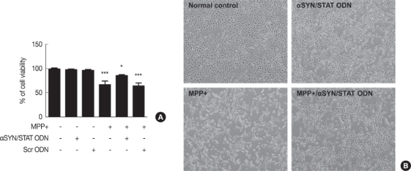
The cytotoxic effects of αSYN/STAT ODN on SH-SY5Y cells were examined through a CCK assay before investigating its pharmacological potential. αSYN/STAT ODN significantly increased the viability of 3 mM MPP+ stimulated SH-SY5Y cells compared to cells treated with only MPP+ (Fig. 1A). Transfection of Scr ODN, a negative control, was similar to cells treated with MPP+. These results were also observed in cell morphology. SH-SY5Y cells grew well, showing obvious neurites, and the cells treated with only αSYN/STAT ODN did not show any difference in cell growth compared to normal cells (Fig. 1B). When SH-SY5Y cells were exposed to MPP+ or Scr ODN, neurites were reduced and cell debris increased; however, they were recovered with αSYN/STAT ODN transfection.
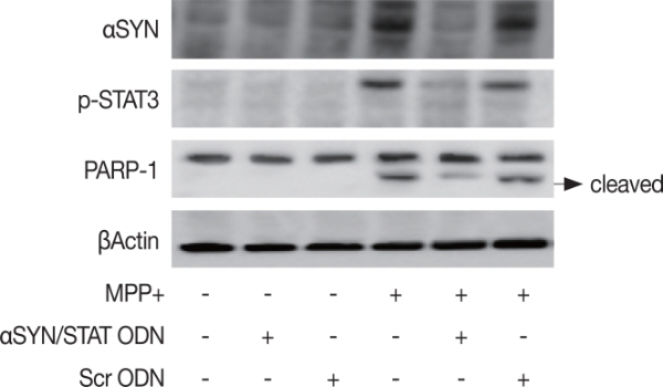
Since apoptosis is one of the important steps in the pathogenesis of PD, we hypothesized that αSYN/STAT ODN could protect dopaminergic neuronal cells by inhibiting the apoptotic pathway. First, we confirmed changes in the expression of ODN target proteins caused by MPP+. As shown Fig. 2, expression of SYN and p-STAT3 were increased by MPP+ or Scr ODN. As expected, this increase was reduced by αSYN/STAT ODN. In addition, αSYN/STAT ODN inhibited MPP+ induced cleaved PARP-1, apoptosis marker protein, in SH-SY5Y cells.
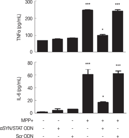
MPP+ causes mitochondrial dysfunction and neuroinflammation [7]. Repression of the JAK/STAT pathway disrupts the neuroinflammation and neurodegeneration circuitry characteristic of PD [8]. To evaluate the impact of αSYN/STAT ODN on MPP+ mediated neuroinflammatory response, SH-SY5Y cells were transfected with αSYN/STAT ODN and Scr ODN followed by MPP+ for 24 hours. The secretion of TNFα and IL6 were significantly inhibited in MPP+ stimulated SH-SY5Y cells by αSYN/STAT ODN transfection (Fig. 3). Scr ODN was similar to cells treated with MPP+.
The first peptide inhibitors of STAT proteins were discovered more than a decade ago, and attempts to target STAT signaling for therapeutic purposes are still ongoing [9]. Aberrant activation of the JAK/STAT pathway contributes to a number of autoimmune and neuroinflammatory diseases [10]. Several studies have illustrated that the novel inflammatory signals namely JAK/STAT, can be activated by LPS, TNF-α, IFN-γ, and IL-6 in the brain [11] and contribute to the pathogenesis of neuroinflammatory diseases [5]. The αSYN accumulation in the brain activated microglial and produced inflammatory cytokines or chemokines through the activation of the JAK/STAT pathway in different models of PD [12]. In addition, neurotoxin MPP+ treatment increased STAT1 expression levels and STAT1 phosphorylation and subsequent apoptosis in cerebellar granule neuron cells [13]. Furthermore, pyridone 6, a JAK inhibitor, reduced interferon β neurotoxicity in SH-SY5Y cells by reducing STAT1 and STAT3 phosphorylation and apoptosis [14].
Our research investigated the αSYN/STAT ODN protective effects on neurotoxicity in SH-SY5Y cells treated by MPP+. Our results exhibited that MPP+ exposure induced neuroinflammatory responses and apoptosis through the secretion of TNFα/IL6 and expression of cleaved PARP-1 in SH-SY5Y cells. In contrast, the transfected αSYN/STAT ODN reversed these changes caused by MPP+ in SH-SY5Y cells. These results strongly support the effectiveness of αSYN/STAT ODN, as the effect of Scr ODN was not observed.
Thus, gene therapy targeted to suppress mRNA level of SYN and transcription activity of STAT simultaneously might provide a new therapeutic strategy to prevent various neurological disorders.